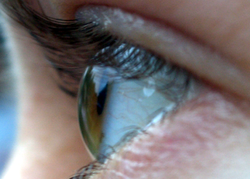
The visual system comprises the sensory organ (the eye) and parts of the central nervous system (the retina containing photoreceptor cells, the optic nerve, the optic tract and the visual cortex) which gives organisms the sense of sight (the ability to detect and process visible light) as well as enabling the formation of several non-image photo response functions. It detects and interprets information from the optical spectrum perceptible to that species to “build a representation” of the surrounding environment. The visual system carries out a number of complex tasks, including the reception of light and the formation of monocular neural representations, colour vision, the neural mechanisms underlying stereopsis and assessment of distances to and between objects, the identification of a particular object of interest, motion perception, the analysis and integration of visual information, pattern recognition, accurate motor coordination under visual guidance, and more. The neuropsychological side of visual information processing is known as visual perception, an abnormality of which is called visual impairment, and a complete absence of which is called blindness. Non-image forming visual functions, independent of visual perception, include (among others) the pupillary light reflex and circadian photoengraving. # ISO certification in India
This article mostly describes the visual system of mammals, humans in particular, although other animals have similar visual systems (see bird vision, vision in fish, mollusc eye, and reptile vision). # ISO certification in India

Mechanical
Together, the cornea and lens refract light into a small image and shine it on the retina. The retina transduces this image into electrical pulses using rods and cones. The optic nerve then carries these pulses through the optic canal. Upon reaching the optic chiasm the nerve fibers decussate (left becomes right). The fibers then branch and terminate in three places.
Neural
Most of the optic nerve fibers end in the lateral geniculate nucleus (LGN). Before the LGN forwards the pulses to V1 of the visual cortex (primary) it gauges the range of objects and tags every major object with a velocity tag. These tags predict object movement. # ISO certification in India
The LGN also sends some fibers to V2 and V3.
V1 performs edge-detection to understand spatial organization (initially, 40 milliseconds in, focusing on even small spatial and color changes. Then, 100 milliseconds in, upon receiving the translated LGN, V2, and V3 info, also begins focusing on global organization). V1 also creates a bottom-up saliency map to guide attention or gaze shift. # ISO certification in India
V2 both forwards (direct and via pulvinar) pulses to V1 and receives them. Pulvinar is responsible for saccade and visual attention. V2 serves much the same function as V1, however, it also handles illusory contours, determining depth by comparing left and right pulses (2D images), and foreground distinguishment. V2 connects to V1 – V5. # ISO certification in India
V3 helps process ‘global motion’ (direction and speed) of objects. V3 connects to V1 (weak), V2, and the inferior temporal cortex.

