V4 recognizes simple shapes, and gets input from V1 (strong), V2, V3, LGN, and pulvinar. V5’s outputs include V4 and its surrounding area, and eye-movement motor cortices (frontal eye-field and lateral intraparietal area).
V5’s functionality is similar to that of the other V’s, however, it integrates local object motion into global motion on a complex level. V6 works in conjunction with V5 on motion analysis. V5 analyzes self-motion, whereas V6 analyzes motion of objects relative to the background. V6’s primary input is V1, with V5 additions. V6 houses the topographical map for vision. V6 outputs to the region directly around it (V6A). V6A has direct connections to arm-moving cortices, including the premotor cortex.

The inferior temporal gyrus recognizes complex shapes, objects, and faces or, in conjunction with the hippocampus, creates new memories. The pretectal area is seven unique nuclei. Anterior, posterior and medial pretectal nuclei inhibit pain (indirectly), aid in REM, and aid the accommodation reflex, respectively.The Edinger-Westphal nucleus moderates pupil dilation and aids (since it provides parasympathetic fibers) in convergence of the eyes and lens adjustment.Nuclei of the optic tract are involved in smooth pursuit eye movement and the accommodation reflex, as well as REM. # ISO certification in India
The suprachiasmatic nucleus is the region of the hypothalamus that halts production of melatonin (indirectly) at first light.
Structure

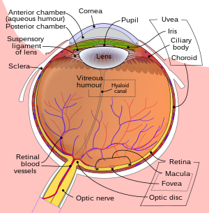
The human eye (horizontal section)
The image projected onto the retina is inverted due to the optics of the eye.
- The eye, especially the retina
- The optic nerve
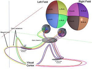
- The optic chiasma
- The optic tract
- The lateral geniculate body
- The optic radiation
- The visual cortex
- The visual association cortex.
These are components of the visual pathway also called the optic pathway that can be divided into anterior and posterior visual pathways. The anterior visual pathway refers to structures involved in vision before the lateral geniculate nucleus. The posterior visual pathway refers to structures after this point.
Eye
Main articles: Eye and Anterior segment of eyeball
Light entering the eye is refracted as it passes through the cornea. It then passes through the pupil (controlled by the iris) and is further refracted by the lens. The cornea and lens act together as a compound lens to project an inverted image onto the retina.
Retina

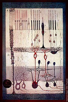
S. Ramón y Cajal, Structure of the Mammalian Retina, 1900
Main article: Retina
The retina consists of many photoreceptor cells which contain particular protein molecules called opsins. In humans, two types of opsins are involved in conscious vision: rod opsins and cone opsins. (A third type, melanopsin in some retinal ganglion cells (RGC), part of the body clock mechanism, is probably not involved in conscious vision, as these RGC do not project to the lateral geniculate nucleus but to the pretectal olivary nucleus.) An opsin absorbs a photon (a particle of light) and transmits a signal to the cell through a signal transduction pathway, resulting in hyper-polarization of the photoreceptor.
Rods and cones differ in function. Rods are found primarily in the periphery of the retina and are used to see at low levels of light. Cones are found primarily in the center (or fovea) of the retina.There are three types of cones that differ in the wavelengths of light they absorb; they are usually called short or blue, middle or green, and long or red. Cones are used primarily to distinguish color and other features of the visual world at normal levels of light. # ISO certification in India
In the retina, the photoreceptors synapse directly onto bipolar cells, which in turn synapse onto ganglion cells of the outermost layer, which will then conduct action potentials to the brain. A significant amount of visual processing arises from the patterns of communication between neurons in the retina. About 130 million photo-receptors absorb light, yet roughly 1.2 million axons of ganglion cells transmit information from the retina to the brain. The processing in the retina includes the formation of center-surround receptive fields of bipolar and ganglion cells in the retina, as well as convergence and divergence from photoreceptor to bipolar cell. In addition, other neurons in the retina, particularly horizontal and amacrine cells, transmit information laterally (from a neuron in one layer to an adjacent neuron in the same layer), resulting in more complex receptive fields that can be either indifferent to color and sensitive to motion or sensitive to color and indifferent to motion. # ISO certification in India

