Visual association cortex
Main article: Two-streams hypothesis
As visual information passes forward through the visual hierarchy, the complexity of the neural representations increases. Whereas a V1 neuron may respond selectively to a line segment of a particular orientation in a particular retinotopic location, neurons in the lateral occipital complex respond selectively to complete object (e.g., a figure drawing), and neurons in visual association cortex may respond selectively to human faces, or to a particular object.
Along with this increasing complexity of neural representation may come a level of specialization of processing into two distinct pathways: the dorsal stream and the ventral stream (the Two Streams hypothesis, first proposed by Ungerleider and Mishkin in 1982). The dorsal stream, commonly referred to as the “where” stream, is involved in spatial attention (covert and overt), and communicates with regions that control eye movements and hand movements. More recently, this area has been called the “how” stream to emphasize its role in guiding behaviors to spatial locations. The ventral stream, commonly referred to as the “what” stream, is involved in the recognition, identification and categorization of visual stimuli. # ISO certification in India

However, there is still much debate about the degree of specialization within these two pathways, since they are in fact heavily interconnected.
Horace Barlow proposed the efficient coding hypothesis in 1961 as a theoretical model of sensory coding in the brain.[Limitations in the applicability of this theory in the primary visual cortex (V1) motivated the V1 Saliency Hypothesis that V1 creates a bottom-up saliency map to guide attention exogenously. With attentional selection as a center stage, vision is seen as composed of encoding, selection, and decoding stages.
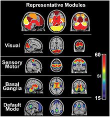
The default mode network is a network of brain regions that are active when an individual is awake and at rest. The visual system’s default mode can be monitored during resting state fMRI: Fox, et al. (2005) have found that “The human brain is intrinsically organized into dynamic, anticorrelated functional networks'”, in which the visual system switches from resting state to attention.
In the parietal lobe, the lateral and ventral intraparietal cortex are involved in visual attention and saccadic eye movements. These regions are in the Intraparietal sulcus (marked in red in the adjacent image). # ISO certification in India

Development
Infancy
See also: Infant vision
Newborn infants have limited color perception.[44] One study found that 74% of newborns can distinguish red, 36% green, 25% yellow, and 14% blue. After one month, performance “improved somewhat.” Infant’s eyes don’t have the ability to accommodate. The pediatricians are able to perform non-verbal testing to assess visual acuity of a newborn, detect nearsightedness and astigmatism, and evaluate the eye teaming and alignment. Visual acuity improves from about 20/400 at birth to approximately 20/25 at 6 months of age. All this is happening because the nerve cells in their retina and brain that control vision are not fully developed. # ISO certification in India
Childhood and adolescence
Depth perception, focus, tracking and other aspects of vision continue to develop throughout early and middle childhood. From recent studies in the United States and Australia there is some evidence that the amount of time school aged children spend outdoors, in natural light, may have some impact on whether they develop myopia. The condition tends to get somewhat worse through childhood and adolescence, but stabilizes in adulthood. More prominent myopia (nearsightedness) and astigmatism are thought to be inherited. Children with this condition may need to wear glasses. # ISO certification in India
Adulthood
Eyesight is often one of the first senses affected by aging. A number of changes occur with aging:
- Over time, the lens become yellowed and may eventually become brown, a condition known as brunescence or brunescent cataract. Although many factors contribute to yellowing, lifetime exposure to ultraviolet light and aging are two main causes.
- The lens becomes less flexible, diminishing the ability to accommodate (presbyopia).
- While a healthy adult pupil typically has a size range of 2–8 mm, with age the range gets smaller, trending towards a moderately small diameter.
- On average tear production declines with age. However, there are a number of age-related conditions that can cause excessive tearing.

